How to Read Arterial Blood Gas Report
Interpretation of arterial blood gases (ABGs) is a crucial skill that a lot of pupil nurses and medical practitioners demand to learn. In this guide, nosotros'll help you lot understand the concepts behind arterial claret gas and teach you the easiest and nearly fun style to interpret ABGs using the tic-tac-toe method.
What is arterial claret gas?

An arterial blood gas is a laboratory test to monitor the patient'due south acid-base residue. It is used to determine the extent of the compensation by the buffer system and includes the measurements of the acidity (pH), levels of oxygen, and carbon dioxide in arterial blood. Dissimilar other blood samples obtained through a vein, a blood sample from an arterial blood gas (ABG) is taken from an artery (commonly on radial or brachial artery).
What are the components of arterial blood gas?
At that place are half dozen components of arterial claret gas (ABGs):
pH
The pH is the concentration of hydrogen ions and determines the acidity or alkalinity of body fluids. A pH of 7.35 indicates acidosis and a pH greater than vii.45 indicates alkalosis.The normal ABG level for pH is vii.35 to 7.45.
PaCO2 (Partial Pressure of Carbon Dioxide)
PaCO2 or fractional force per unit area of carbon dioxide shows the capability of the gas exchange between the alveoli and the external environment (alveolar ventilation). Carbon dioxide (CO2) cannot escape when there is damage in the alveoli, excess CO2 combines with h2o to course carbonic acid (H2CO3) causing an acidotic state. When there is hypoventilation in the alveolar level (for example, in COPD), the PaCO2 is elevated, and respiratory acidosis results. On the other hand, when in that location is alveolar hyperventilation (e.g., hyperventilation), the PaCO2 is decreased causing respiratory alkalosis. For PaCO2, the normal range is 35 to 45 mmHg (respiratory determinant).
PaO2 (Fractional Pressure of Oxygen)
PaO2 or fractional pressure of oxygen or PAO2 indicates the amount of oxygen available to bind with hemoglobin. The pH plays a role in the combining power of oxygen with hemoglobin: a depression pH means in that location is less oxygen in the hemoglobin. For PaO2, the normal range is 75 to 100 mmHg
SO2 (Oxygen Saturation)
SOtwo or oxygen saturation, measured in percentage, is the amount of oxygen in the blood that combines with hemoglobin. Information technology can be measured indirectly past computing the PAO2 and pH Or measured directly by co-oximetry. Oxygen saturation, the normal range is 94–100%
HCO3 (Bicarbonate)
HCO3 or bicarbonate ion is an alkaline metal substance that comprises over one-half of the total buffer base in the blood. A deficit of bicarbonate and other bases indicates metabolic acidosis. Alternatively, when there is an increment in bicarbonates present, then metabolic alkalosis results.
Be (Base Backlog)
BE. Base excess or BE value is routinely checked with HCOthree value. A base excess of less than –ii is acidosis and greater than +ii is alkalosis. Base backlog, the normal range is –ii to +2 mmol/L
Normal Values in Arterial Blood Gas
To determine acrid-base of operations imbalance, you demand to know and memorize these values to recognize what deviates from normal. The normal range for ABGs is used as a guide, and the decision of disorders is often based on blood pH. If the blood is basic, the HCOthree level is considered considering the kidneys regulate bicarbonate ion levels. If the blood is acidic, the PaCO2 or partial pressure of carbon dioxide in arterial blood is assessed because the lungs regulate the majority of acid. The normal ABG values are the following:
- For pH, the normal range is 7.35 to 7.45
- For PaCOii, the normal range is 35 to 45 mmHg (respiratory determinant)
- For PaO2, the normal range is 75 to 100 mmHg
- For HCO3, the normal range is 22 to 26 mEq/50 (metabolic determinant)
- Oxygen saturation, the normal range is 94–100%
- Base of operations excess, the normal range is –2 to +2 mmol/Fifty
Interpreting Arterial Claret Gas Imbalances
Interpreting arterial blood gases is used to notice respiratory acidosis or alkalosis, or metabolic acidosis or alkalosis during an acute disease. To decide the blazon of arterial blood gas the key components are checked. The best (and fun) mode of interpreting arterial claret gas is by using the tic-tac-toe method below:
Goals of Arterial Claret Gas analysis
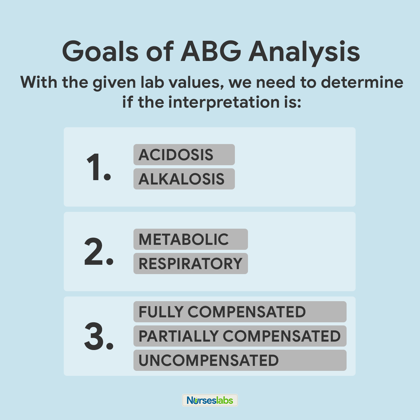
For the purpose of this guide, we accept set three (3) goals that we demand to attain when interpreting arterial blood gases. The goals are as follows:
- Based on the given ABG values, determine if values interpret ACIDOSIS or ALKALOSIS.
- 2d, nosotros need to make up one's mind if values define METABOLIC or RESPIRATORY.
- Lastly, we need to determine the compensation if it is: FULLY COMPENSATED, PARTIALLY COMPENSATED, or UNCOMPENSATED.
We need to keep these goals in mind as they'll come up up later in the steps for the ABG interpretation technique.
Steps in ABG assay using the tic-tac-toe method
At that place are eight (viii) steps unproblematic steps you lot need to know if you want to interpret arterial blood gases (ABGs) results using the tic-tac-toe technique.
1. Memorize the normal values.
The first step is you need to familiarize yourself with the normal and abnormal ABG values when yous review the lab results. They are easy to remember:
- For pH, the normal range is 7.35 to 7.45
- For PaCOii, the normal range is 35 to 45
- For HCO3, the normal range is 22 to 26
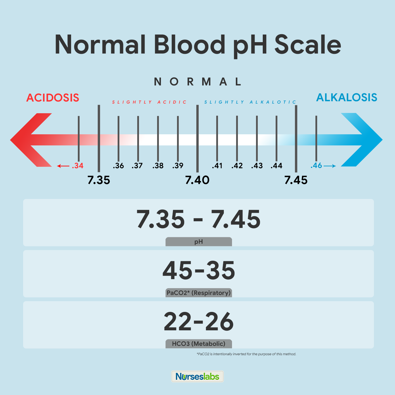
The recommended way of memorizing it is past drawing the diagram of normal values above. Write information technology downward together with the arrows indicating ACIDOSIS or ALKALOSIS. Notation that PaCO2 is intentionally inverted for the purpose of the Tic-Tac-Toe method.
two. Create your tic-tac-toe grid.

Once you lot've memorized the normal values and the diagram, create a blank your tic-tac-toe filigree and characterization the top row equally ACIDOSIS, NORMAL, and ALKALOSIS. Based on their values, we need to decide in which column we'll place pH, PaCO2, and HCO3 in the grid.
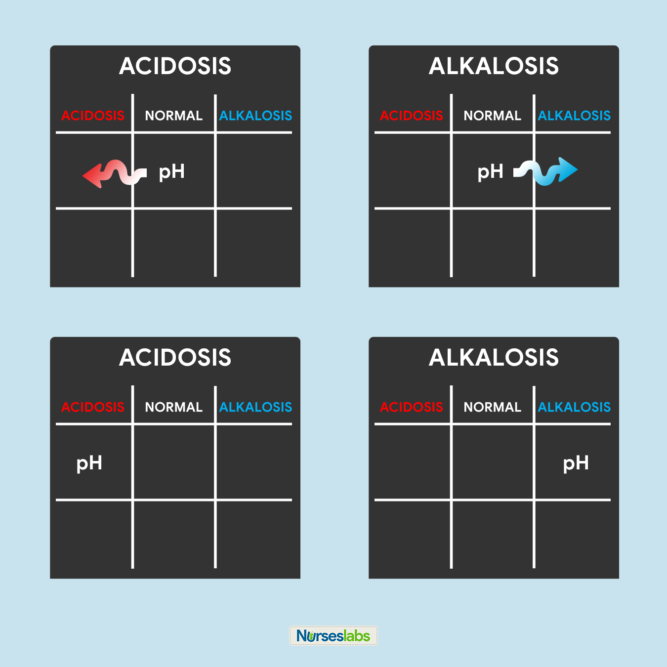
3. Determine if pH is under NORMAL, ACIDOSIS, or ALKALOSIS.
The tertiary footstep of this technique is to make up one's mind the acidity or alkalinity of the blood with the given value of the pH equally our determining factor. Remember in stride #1 that the normal pH range is from 7.35 to 7.45.
- If the blood pH is between vii.35 to seven.39, the interpretation is NORMAL simply SLIGHTLY ACIDOSIS, place it nether the NORMAL column.
- If the blood pH is between vii.41 to 7.45, interpretation is NORMAL merely SLIGHTLY ALKALOSIS, place it under the NORMAL column.
- Any blood pH beneath 7.35 (vii.34, seven.33, vii.32, and so on…) is ACIDOSIS, place information technology under the ACIDOSIS column.
- Any claret pH to a higher place seven.45 (7.46, 7.47, 7.48, and and so on…) is ALKALOSIS, place it under the ALKALOSIS column.
Please utilize the diagram below to assistance you visualize whether the normal value is ACIDOSIS or ALKALOSIS.
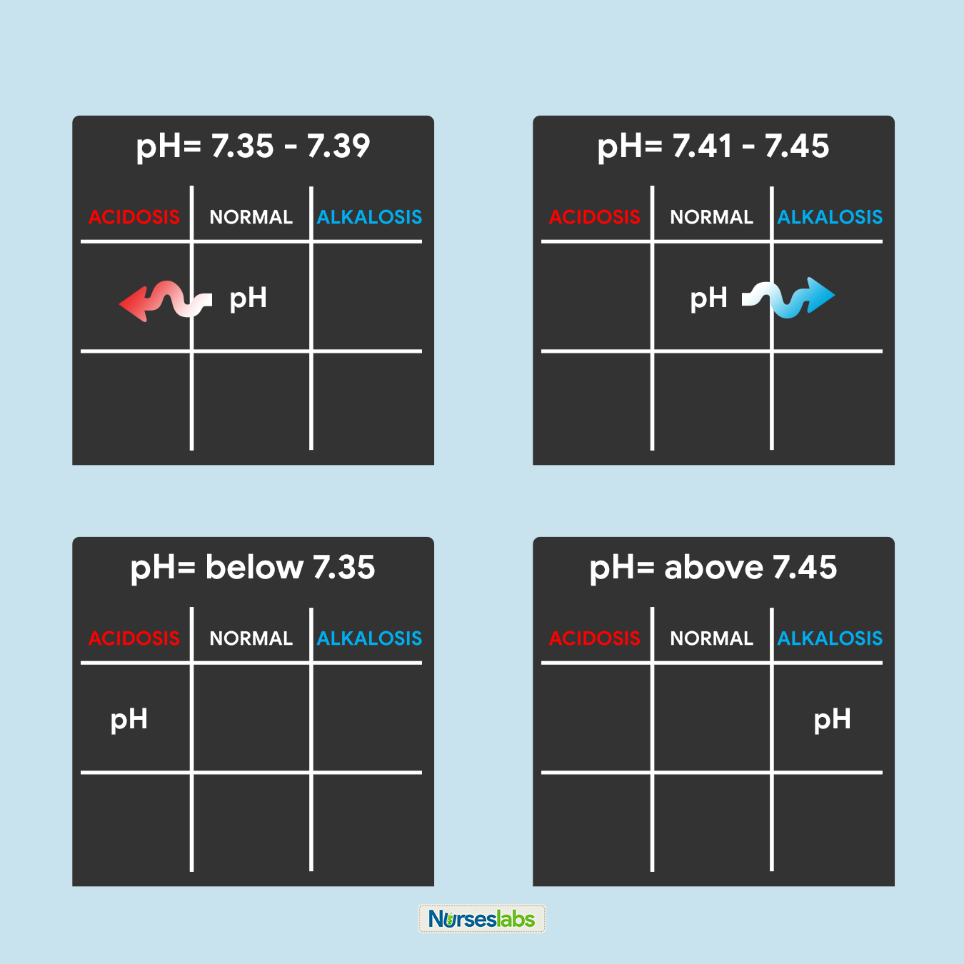
Once you've determined whether the pH is nether the ACIDOSIS or ALKALOSIS, plot it on your tic-tac-toe grid under the advisable column.
4. Determine if PaCO2 is under NORMAL, ACIDOSIS, or ALKALOSIS.
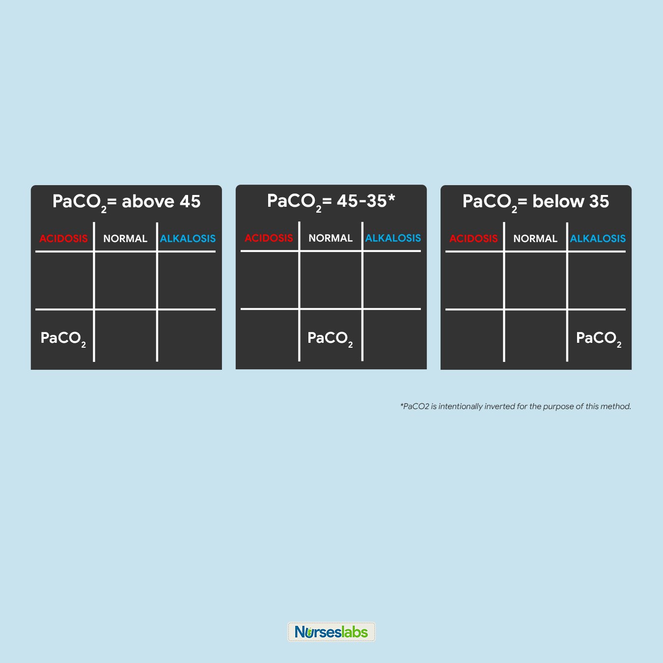
For this step, we need to interpret if the value of PaCO2 is inside the NORMAL range, ACIDIC, or BASIC and plot it on the grid under the appropriate column. Retrieve that the normal range for PaCO2 is from 35 to 45:
- If PaCOii is below 35, place information technology under the ALKALOSIS cavalcade.
- If PaCO2 is above 45, place it under the ACIDOSIS column.
- If PaCO2 is inside its normal range, identify it under the NORMAL column.
5. Make up one's mind if HCOthree is under NORMAL, ACIDOSIS, or ALKALOSIS.

Next, nosotros need to interpret if the value of HCO3 is within the NORMAL range, ACIDIC, or Bones and plot information technology under the appropriate column in the tic-tac-toe grid. Remember that the normal range for HCO3 is from 22 to 26:
- If HCO3 is below 22, place it under the ACIDOSIS column.
- If HCO3 is above 26, place it under the ALKALOSIS column.
- If HCOiii is within its normal range, place it under the NORMAL column.
6. Solve for goal #one: ACIDOSIS or ALKALOSIS.
Now, we will start solving for our goals. Looking at the tic-tac-toe grid, decide whether in what column the pH is placed and translate the results:
- If pH is under the ACIDOSIS column, it is ACIDOSIS.
- If pH is nether the ALKALOSIS column, it is ALKALOSIS.
- If pH is under the NORMAL column, determine whether the value is leaning towards ACIDOSIS or ALKALOSIS and interpret accordingly.
In this step, nosotros tin reach goal #1 of determining ACIDOSIS or ALKALOSIS.
vii. Solve for goal #2: METABOLIC or RESPIRATORY.

Looking back again on the tic-tac-toe grid, decide if pH is under the same column every bit PaCO2 or HCOthree then nosotros tin can accomplish our goal #ii of determining if the ABG is RESPIRATORY or METABOLIC. Interpret the results as follows:
- If pH is nether the same column as PaCO2, it is RESPIRATORY.
- If pH is under the same cavalcade as HCOthree, it is METABOLIC.
- If pH is under the NORMAL column, determine whether the value is leaning towards ACIDOSIS or ALKALOSIS and translate accordingly.
eight. Solve for goal #iii: COMPENSATION.
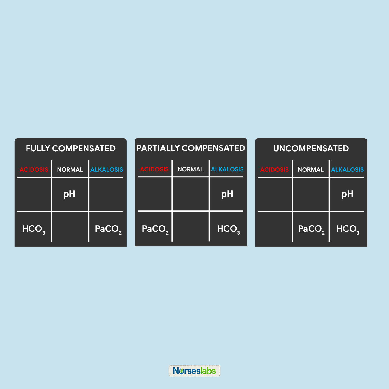
Lastly, we need to decide the compensation to accomplish our goal #3. Interpret the results as follows:
- It is FULLY COMPENSATED if pH is normal.
- It is PARTIALLY COMPENSATED if all three (iii) values are aberrant.
- It is UNCOMPENSATED if PaCO2 or HCOthree is normal and the other is abnormal.
Application and Examples
Allow'due south solve for the ABG interpretation with the examples beneath:
Practice Problem #1:
pH=7.26 | PaCOii=32 | HCO3=xviii
- Think the normal values.
- Brand your tic-tac-toe grid.
- pH of 7.26 ABNORMAL and under ACIDOSIS, so we identify pH under ACIDOSIS.
- PaCOii of 32 is ABNORMAL and under ALKALOSIS, so we place PaCO2 under ALKALOSIS.
- HCO3 of eighteen is Abnormal and under ACIDOSIS, so we place HCO3 under ACIDOSIS.
- pH is under ACIDOSIS, therefore solving for goal #ane, we accept ACIDOSIS.
- pH is on the same cavalcade as HCO3, therefore solving for goal #ii, nosotros have METABOLIC.
- All three values are ABNORMAL, therefore solving for goal #iii, we have a PARTIALLY COMPENSATED ABG.
The answer to Practice Problem #1:
Metabolic Acidosis, Partially Compensated
Practise Trouble #2:
pH=7.44 | PaCO2=thirty | HCO3=21
- Remember the normal values.
- Make your tic-tac-toe grid.
- pH of vii.44 is NORMAL but slightly leaning towards ALKALOSIS, so we identify pH nether the NORMAL cavalcade with an pointer pointing towards the ALKALOSIS cavalcade.
- PaCO2 of thirty is ABNORMAL and ALKALOSIS, so we place PaCOii under the ALKALOSIS column.
- HCO3 of 21 is ABNORMAL and ACIDOSIS, and so we place HCO3 under the ACIDOSIS column.
- pH of 7.44 is NORMAL just leaning towards ALKALOSIS, therefore solving for goal #1, nosotros have ALKALOSIS.
- pH is NORMAL but is leaning towards ALKALOSIS, therefore under the aforementioned column as PaCOtwo. Solving for goal #ii, we have RESPIRATORY.
- pH is NORMAL, therefore solving for goal #iii, we have a FULLY COMPENSATED ABG.
The answer to Practice Problem #2:
Respiratory Alkalosis, Fully Compensated
Exercise Problem #3:
pH=seven.1 | PaCO2=xl | HCO3=eighteen
- Recollect the normal values.
- Make your tic-tac-toe grid.
- pH of 7.one is ABNORMAL and ACIDOSIS, therefore, we place pH under the ACIDOSIS cavalcade in the tic-tac-toe filigree.
- PaCO2 of 40 is NORMAL, therefore, place it nether the NORMAL column.
- HCOthree of 18 is Abnormal and ACIDOSIS, so we place HCOiii nether the ACIDOSIS column.
- pH of 7.one is ACIDOSIS, therefore, solving for goal #1, we have ACIDOSIS.
- pH is under the same column equally HCOiii, therefore, solving for goal #2, we have determined that it is METABOLIC.
- pH is Aberrant then as HCO3, just PaCO3 is under the NORMAL cavalcade. Solving for goal #3, we can interpret it equally UNCOMPENSATED.
The answer to Practice Problem #iii:
Metabolic Acidosis, Uncompensated
How to depict Arterial Blood Gas?
Arterial claret is usually fatigued via the brachial or radial artery.
- Inform that client about the procedure and that in that location is no food or fluid restriction imposed.
- Notation if the client is taking anticoagulant therapy or aspirin every bit this may touch results.
- Annotation if the client is receiving oxygen therapy (flow rate, type of administration device), and the client's electric current temperature.
- Using a heparinized needle and syringe, collect one to v mL of arterial blood. Common sites for drawing arterial claret are the radial and brachial artery.
- Put the syringe with arterial blood in an water ice-h2o bag to minimize the metabolic action of the sample.
- Evangelize the blood sample immediately to the laboratory.
- Utilise pressure to the puncture site for 5 minutes or longer.
Acid-Base Balance and Imbalances
Acrid-base of operations imbalances develop when a person's normal homeostatic mechanisms are dysfunctional or overwhelmed. I type of acid-base of operations imbalance is acidosis wherein the blood is relatively too acidic (low pH). The body produces two types of acrid, therefore, there are ii types of acidosis: respiratory acidosis and metabolic acidosis. On the contrary, alkalosis is a condition wherein the blood is relatively as well basic (loftier pH), there are also two types of alkalosis: respiratory alkalosis and metabolic alkalosis.
When acid-base of operations imbalances occur, the body activates its compensatory mechanisms (the lungs and kidneys) to help normalize the claret pH. The kidneys compensate for respiratory acid-base imbalances while the respiratory organisation compensates for metabolic acid-base imbalances. This does not correct the root cause of the trouble, if the underlying condition is not corrected, these systems will neglect.
Respiratory Acidosis
Respiratory acidosis occurs when breathing is inadequate (alveolar hypoventilation) and the lungs are unable to excrete enough CO2 causing PaCO2 or respiratory acid builds upwardly. The extra CO2 combines with water to form carbonic acid, causing a state of acidosis — a mutual occurrence in emphysema. The kidneys actuate its compensatory process (albeit dull, oftentimes 24 hours or more than) by increasing the excretion of metabolic acids through urination, which increases blood bicarbonate.
Types of Respiratory Acidosis
There are two forms of respiratory acidosis: Acute and Chronic.
- Acute respiratory acidosis. This class of respiratory acidosis occurs immediately. Left untreated, symptoms will go progressively worse. It'southward a medical emergency and can become life-threatening.
- Chronic respiratory acidosis. This grade of respiratory acidosis develops through time. It doesn't cause symptoms. Instead, the body adapts to the increased acidity. For case, the kidneys produce more than bicarbonate to help maintain residuum. Chronic respiratory acidosis may not cause symptoms. Developing some other illness may cause chronic respiratory acidosis to worsen and become astute respiratory acidosis.
Risk Factors
Respiratory acidosis is typically caused past an underlying disease or condition. This is as well called respiratory failure or ventilatory failure.
- Hypoventilation. A decrease in ventilation increases the concentration of carbon dioxide in the blood and decreases the blood's pH (brain trauma, coma, hypothyroidism: myxedema).
- Chronic Obstructive Pulmonary Disease (COPD). In chronic respiratory acidosis in COPD patients, the body tries to compensate by retaining more than bicarbonate to overcome acidosis.
- Respiratory Conditions. The lungs are not able to eliminate enough of the carbon dioxide produced by the body. Excess carbon dioxide causes the pH of the blood and other actual fluids to subtract, making them besides acidic. (pneumothorax, pneumonia, status asthmaticus)
- Drug Intake. Overdose of an opiate or opioid, such as morphine, tramadol, heroin, fentanyl, or magnesium sulfate (MgSO4) tin cause respiratory acidosis.
Signs and Symptoms
Signs and symptoms of respiratory acidosis are every bit follows:
- Altered level of consciousness. Respiratory acidosis may be the result of an altered level of consciousness caused by encephalopathy or cerebral edema.
- Confusion. Acute respiratory acidosis may besides cause symptoms involving the brain, including confusion, stupor, drowsiness, and muscle jerks.
- Disorientation. Respiratory acidosis may result in disorientation, headache, or even focal neurologic signs.
- Blackout. When the lungs can't remove all of the carbon dioxide produced by the body through normal metabolism, the claret becomes acidified, leading to increasingly serious symptoms, from sleepiness to coma.
- Tremors. Manifest as shaking or jerking muscle movements.
- Asterixis. An inability to maintain the posture of part of the trunk.
Management of Respiratory Acidosis
Medical and nursing management of an arterial claret gas of respiratory acidosis includes the post-obit:
- Treat underlying atmospheric condition.
- Medications. Bronchodilator medicines and corticosteroids may be used to contrary some types of airway obstacle, like those linked to asthma and COPD.
- Weight loss. In the case of obesity hypoventilation syndrome, significant weight loss may exist necessary to reduce abnormal compression of the lungs.
- Provide mechanical ventilation through oxygen supplementation. Boosted oxygen may be provided to convalesce the depression oxygen level in the blood.
- Manage hyperkalemia through the use of Kayexalate. Acidosis causes potassium to move from cells to extracellular fluid (plasma) in exchange for hydrogen ions, and alkalosis causes the reverse motion of potassium and hydrogen ions. Kayexalate increases fecal potassium excretion through the bounden of potassium in the lumen of the gastrointestinal tract.
- Maintain adequate hydration. Provide intravenous fluids and electrolytes as ordered.
Respiratory Alkalosis
Respiratory alkalosis can result from hyperventilation since the lungs excrete too much carbonic acrid which increases pH. Since respiratory alkalosis occurs quickly, the kidneys practice not have time to compensate. Neurological symptoms such as confusion, paresthesias, and cell membrane excitability occur when the claret pH, CSF, and ICF increases acutely.
Risk Factors
Causes of hyperventilation include:
- Panic. Panic attacks and anxiety are the most common causes of hyperventilation.
- Hyperthermia. Fever may manifest as hyperventilation. The exact mechanism is not known merely is thought to be due to carotid body or hypothalamic stimulation by the increased temperature.
- Brainstem damage. Central neurogenic hyperventilation (CNH) is the human trunk'southward response to reduced carbon dioxide levels in the blood. This reduction in carbon dioxide is acquired past the contraction of cranial arteries from harm acquired by lesions in the brain stem.
- Metabolic acidosis. Hyperventilation occurs virtually often as a response to hypoxia, metabolic acidosis, increased metabolic demands, pain, or anxiety.
- Diabetic ketoacidosis (DKA). The simply known compensatory response to metabolic acidosis in DKA is hyperventilation with consecutive respiratory alkalosis.
- Pregnancy. Progesterone levels are increased during pregnancy. Progesterone causes stimulation of the respiratory center, which can lead to respiratory alkalosis.
- Salicylate toxicity. Salicylate toxicity causes respiratory alkalosis and, by an independent mechanism, metabolic acidosis.
Signs and Symptoms
Hyperventilation is a sign that respiratory alkalosis is about likely to occur. All the same, depression carbon dioxide levels in the blood as well have a number of physical effects, including:
- Numbness. Increased neuromuscular irritability in which a person loses feeling in a item part of their trunk.
- Tingling sensation. Prickling sensation that is usually felt in the hands, arms, legs, or feet, just can also occur in other parts of the body.
- Palpitations. Palpitations are the perceived abnormality of the heartbeat characterized by awareness of cardiac muscle contractions in the chest.
- Tetany. Tetany or tetanic seizure is a medical sign consisting of the involuntary contraction of muscles.
- Convulsions. A medical condition where body muscles contract and relax rapidly and repeatedly, resulting in uncontrolled actions of the body.
- Signs and symptoms of hypokalemia and hypocalcemia. Persistent respiratory alkalosis tin can induce secondary hypocalcemia and hypokalemia that may crusade cardiac arrhythmias, conduction abnormalities, and various somatic symptoms such as paresthesia, hyperreflexia, convulsive disorders, muscle spasm, muscle twitching, positive Chvostek's sign, and tetany.
Management of Respiratory Alkalosis
The treatment for respiratory alkalosis depends on the underlying cause. Treating the status is a matter of rising carbon dioxide levels in the blood. The following strategies and tips are useful for respiratory alkalosis acquired by over-breathing due to panic and anxiety.
- Breathe into a paper bag. Animate through a paper purse fills it with carbon dioxide helping in inhaling exhaled air dorsum into the lungs.
- Treat underlying condition:
- Medications. Administering an opioid pain reliever or anti-anxiety medication to reduce hyperventilation.
- Relaxation techniques. Breathing exercises that assist relax and breathe from the diaphragm and abdomen, rather than breast wall.
- Safe. Stay with the patient.
- Lavage. Later on massive aspirin ingestions, aggressive gut decontamination is advisable, including gastric lavage.
- Correction of hypokalemia and hypocalcemia.
- Oxygenation as indicated. Providing oxygen to help continue a person from hyperventilating.
Metabolic Acidosis
Metabolic acidosis is when there is a decrease in bicarbonates and a buildup of lactic acrid occurs. This happens in diarrhea, ketosis, and kidney disorders. It has three master root causes: increased acid production, loss of bicarbonate, and a reduced ability of the kidneys to excrete excess acids.
Risk Factors
- Diabetic Ketoacidosis (DKA). DKA develops when substances chosen ketone bodies (which are acidic) build upwards during uncontrolled diabetes. DKA occurs by and large in Type 1 Diabetes Mellitus (DM).
- Chronic Renal Failure (CRF). This is due to reduced tubular bicarbonate reabsorption and insufficient renal bicarbonate production in relation to the number of acids synthesized past the body and ingested with food.
- Chronic Hypoxia. With chronic hypoxia, metabolic and hypercapnic acidosis develop along with considerable lactate germination and pH falling to beneath half-dozen.8.
- Obesity. Obesity, specially in conjunction with insulin resistance, tin can increase metabolic acidosis and thus upshot in a reduction of urinary citrate excretion.
- Diarrhea. Loss of bicarbonate stores through diarrhea or renal tubular wasting leads to a metabolic acidosis state characterized by increased plasma chloride concentration and decreased plasma bicarbonate concentration.
- Dehydration. Electrolyte disturbances acquired by prolonged airsickness or astringent dehydration can cause metabolic acidosis.
- Aspirin Toxicity. Aspirin overdose causes the body to non produce ATP, leading to anaerobic metabolism with consequent raised lactate and ketone bodies. Acute aspirin or salicylates overdose or poisoning tin can cause initial respiratory alkalosis through metabolic acidosis ensues thereafter.
- Methanol Poisoning. Significant methanol ingestion leads to metabolic acidosis, which is manifested by a low serum bicarbonate level. The anion gap is increased secondary to loftier lactate and ketone levels. This is probably due to formic acid accumulation.
Signs and Symptoms
- Altered level of consciousness
- Confusion
- Disorientation
- Lack of appetite
- Coma
- Jaundice
Management of Metabolic Acidosis
Patients with arterial claret gas indicating metabolic acidosis are managed and treated by:
- Sodium bicarbonate. Indicated in the treatment of metabolic acidosis which may occur in severe renal disease, uncontrolled diabetes, circulatory insufficiency due to daze or severe dehydration, extracorporeal circulation of blood, cardiac arrest, and severe primary lactic acidosis.
- Treat the underlying condition.
- Hydration for diabetic ketoacidosis. The major treatment of this condition is the initial rehydration.
- Dialysis for chronic renal failure. The command of metabolic acidosis in hemodialysis is mainly focused on the supply of bicarbonate during the dialysis sessions.
- Employ of diuretics.
- Initiate safety measures.
- Kayexalate. Acidosis causes potassium to movement from cells to extracellular fluid (plasma) in substitution for hydrogen ions, and alkalosis causes the reverse move of potassium and hydrogen ions. Kayexalate increases fecal potassium excretion through the bounden of potassium in the lumen of the alimentary canal.
Metabolic Alkalosis
Metabolic alkalosis occurs when bicarbonate ion concentration increases, causing an tiptop in blood pH. This can occur in excessive vomiting, aridity, or endocrine disorders.
Run a risk Factors
- Vomiting. Vomiting causes metabolic alkalosis by the loss of gastric secretions, which are rich in hydrochloric acid (HCl). Whenever a hydrogen ion is excreted, a bicarbonate ion is gained in the extracellular space.
- Sodium bicarbonate overdose. Assistants of sodium bicarbonate in amounts that exceed the capacity of the kidneys to excrete this excess bicarbonate may cause metabolic alkalosis.
- Hypokalemia. Due to a low extracellular potassium concentration, potassium shifts out of the cells. In club to maintain electric neutrality, hydrogen shifts into the cells, raising blood pH.
- Nasogastric suction. Just similar in airsickness, nasogastric (NG) suction as well generates metabolic alkalosis past the loss of gastric secretions, which are rich in hydrochloric acrid (HCl).
Signs and Symptoms
Metabolic alkalosis may not prove any symptoms. People with this type of alkalosis more than often mutter of the underlying conditions that are causing it. These can include:
- Numbness
- Vomiting
- Diarrhea
- Swelling in the lower legs (peripheral edema)
- Fatigue
- Tingling sensation
- Agitation
- Disorientation
- Seizures
- Coma
Direction of Metabolic Alkalosis
- Antiemetic. In the case of vomiting, administer antiemetics, if possible.
- Ammonium chloride. Ammonium chloride is a systemic and urinary acidifying amanuensis that is converted to ammonia and hydrochloric acid through oxidation by the liver. Intravenous (IV) ammonium chloride is a handling option for severe cases of metabolic alkalosis.
- Acetazolamide (Diamox). Acetazolamide besides appears to be prophylactic and constructive in patients with metabolic alkalosis post-obit handling of respiratory acidosis from exacerbations of chronic obstructive pulmonary illness (COPD).
Arterial Claret Gas Interpretation Quiz
If you need to do your new skills acquired here, cheque out our Arterial Claret Gas Interpretation for NCLEX (40 Questions)
References and Sources
The following sources are used every bit references for this guide. You may find them interesting for your additional reading:
- Barnette, L., & Kautz, D. D. (2013). Creative means to teach arterial claret gas estimation.Dimensions of Disquisitional Care Nursing,32(ii), 84-87.
- Samuel, R. (2018). A Graphical Tool for Arterial Blood Gas Estimation using Standard Bicarbonate and Base Excess.Indian J Med Biochem,22(1), 85-89.
- Sood, P., Paul, Yard., & Puri, South. (2010). Interpretation of arterial claret gas.Indian journal of critical intendance medicine: peer-reviewed, official publication of Indian Lodge of Disquisitional Care Medicine,14(2), 57.
- Williams, A. J. (1998). Assessing and interpreting arterial blood gases and acrid-base balance.Bmj,317(7167), 1213-1216.
- Verma, A. K., & Roach, P. (2010). The estimation of arterial blood gases.Aust Prescr,33(4), 124-129.
carrasquillodrefoonew.blogspot.com
Source: https://nurseslabs.com/arterial-blood-gas-abgs-interpretation-guide/
0 Response to "How to Read Arterial Blood Gas Report"
Post a Comment